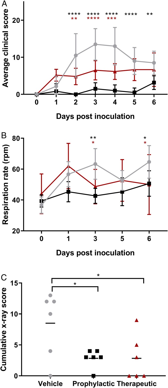
Fig. 2.
Clinical findings in rhesus macaques inoculated with MERS-CoV and treated with remdesivir. Three groups of six rhesus macaques were inoculated with MERS-CoV strain HCoV-EMC/2012; one group was i.v.-administered 1 mL/kg vehicle solution (vehicle control; gray circles), one group was administered 5 mg/kg remdesivir starting at 24 h before inoculation (prophylactic remdesivir; black squares), and one group was administered 5 mg/kg remdesivir starting at 12 h after inoculation (therapeutic remdesivir; red triangles). After inoculation, the animals were observed twice daily for clinical signs of disease and scored using a predetermined clinical scoring system (A). On 0, 1, 3, 5 and 6 dpi, clinical examinations were performed during which respiration rate was determined (B), and radiographs were taken. Radiographs were used to score individual lung lobes for severity of pulmonary infiltrates by a clinical veterinarian according to a standard scoring system (0: normal; 1: mild interstitial pulmonary infiltrates; 2: moderate pulmonary infiltrates perhaps with partial cardiac border effacement and small areas of pulmonary consolidation; 3: serious interstitial infiltrates, alveolar patterns and air bronchograms); the cumulative X-ray score is the sum of the scores of the six individual lung lobes per animal; scores shown are from 6 dpi (C). Asterisks indicate statistically significant difference in a two-way (A and B) or one-way (C) ANOVA with Dunnett’s multiple comparisons; black asterisks indicate statistical significance between the vehicle control and prophylactic remdesivir groups, and red asterisks indicate statistical significance between the vehicle control and therapeutic remdesivir groups. *P < 0.05; **P < 0.01; ***P < 0.001; ****P < 0.0001.

