Fig. 3.
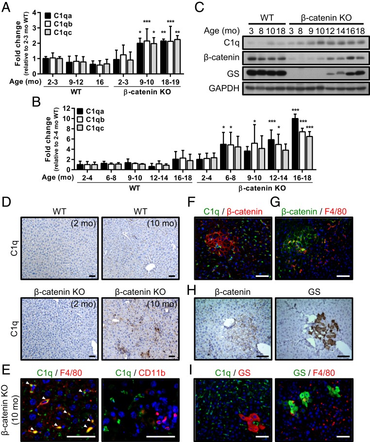
Complement C1q levels are elevated in the inflammatory niche and associated with activation of the β-catenin pathway in periportal HPCs. (A) Results of the microarray analysis showing the levels of the C1qa, C1qb, and C1qc mRNAs in liver tissues collected from WT control and β-catenin KO mice at the indicated ages. The levels were compared with the 2- to 3-mo-old WT livers, in which the level was set to 1. n = 3 to 6 per group. (B and C) qRT-PCR data (B) and representative Western blots (C) showing C1q expression in the liver tissues of WT and β-catenin KO mice at various ages. Levels of C1qa, C1qb, and C1qc mRNA were compared between the β-catenin KO livers and WT livers at 2 to 4 mo. Results are presented as mean ± SD. *P < 0.05; **P < 0.01; ***P < 0.001. n = 3 to 5 per group. (D) IHC staining with C1q antibody in the liver tissues collected from 2- and 10-mo-old WT and β-catenin KO mice. (E) IF staining for C1q, F4/80, and CD11b in liver tissues of 10-mo-old β-catenin KO mice. White arrowheads indicate the colocalization of C1q and F4/80. (F–I) Representative IF and IHC staining with indicated antibodies in 10- to 12-mo-old β-catenin KO liver tissues. (Scale bars: 50 µm.)

