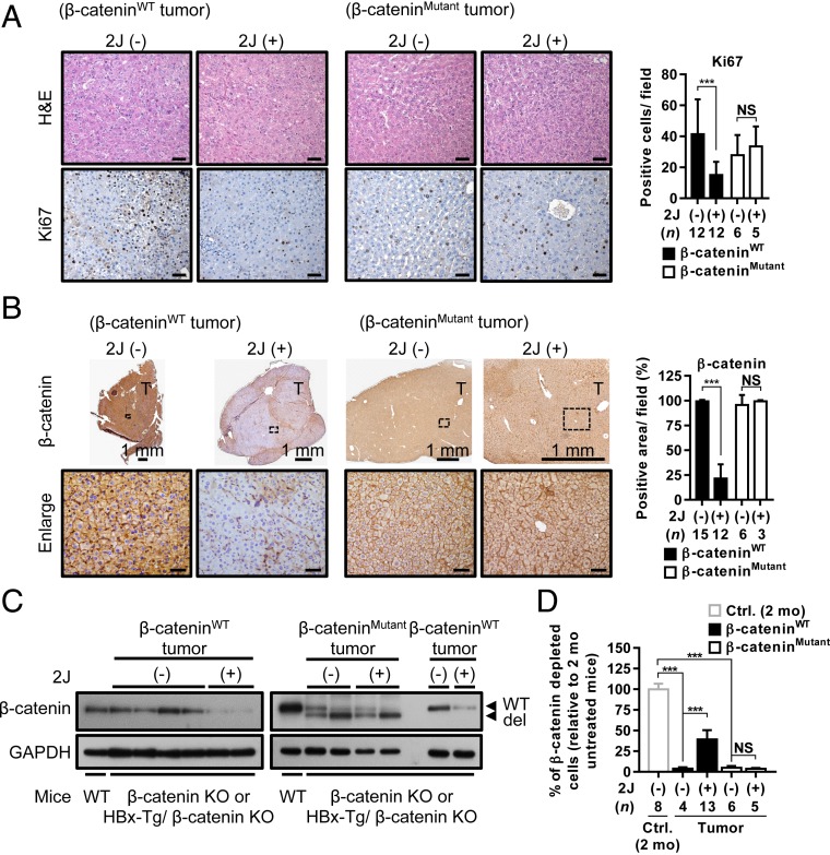
Fig. 5.
Liver tumors containing WT β-catenin are responsive to C1q inhibitor treatment. (A and B) Representative images of H&E and IHC staining for Ki67 (A) and β-catenin (B) in WT and mutant β-catenin–containing liver tumors from β-catenin KO and HBx-Tg/β-catenin KO mice that were not treated or treated with 2J peptide for 2 mo. Percentages of positive signals for Ki67 and β-catenin were quantified and are shown as bar graphs to the right of the images. (C) Western blots showing the expression of β-catenin in WT and mutant β-catenin–containing liver tumors collected from untreated or 2J peptide-treated tumor-bearing mice. β-catenin expression in the liver tissues of WT control mice served as a positive control. (D) Genomic DNA extracted from the liver tissues collected from different groups of mice as indicated were processed for qRT-PCR analysis to evaluate the percentage of cells in which β-catenin was deleted in each liver tissue. The percentage of β-catenin depletion in the liver tissues from 2-mo-old β-catenin KO mice was set to 100%. The β-catenin mutations identified in the tumors were mutations or deletions at around exon 3 of the CTNNB1 gene. Results are presented as mean ± SD. N.S., not significant. ***P < 0.001. (Scale bar: 50 µm for A and B.)

