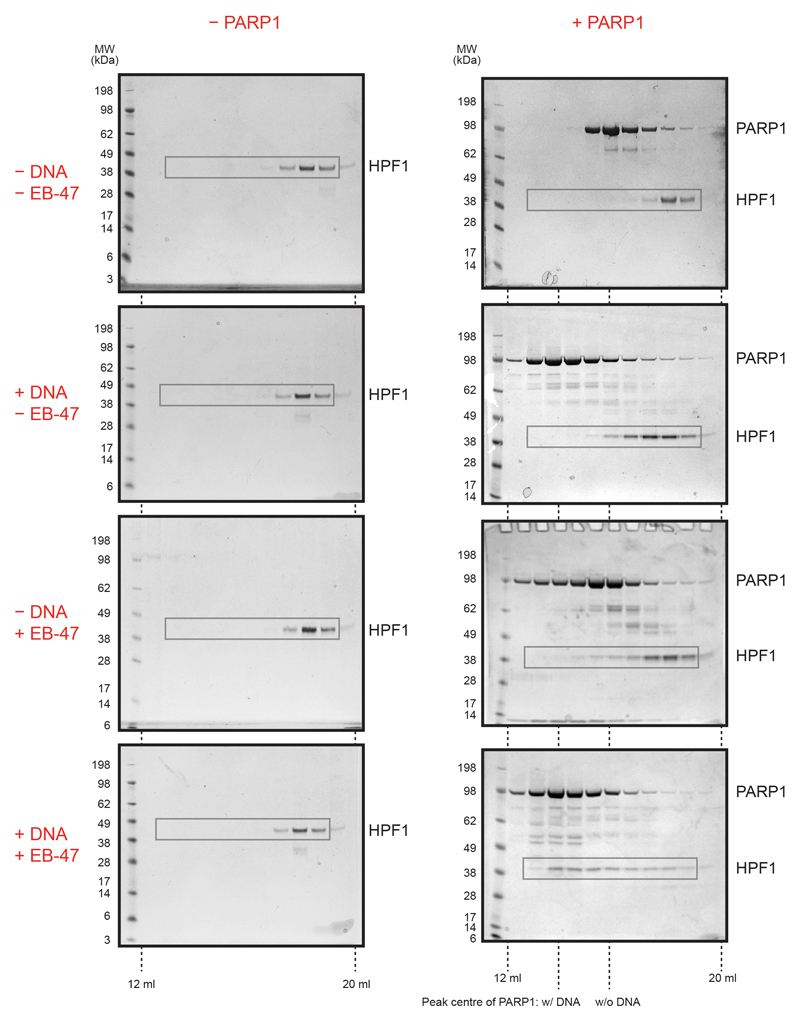
Extended Data Figure 2. Analytical size-exclusion chromatography analysis of HPF1-PARP1 interaction.
Uncropped SDS-PAGE gels with fractions from analytical size-exclusion chromatography. HPF1 was analysed in the presence or absence of PARP1 and either alone or with a short DNA duplex and/or the NAD+ analogue EB-47. Images from Fig. 1d are identical with areas marked with grey rectangles. Note the changed elution profile of PARP1 itself in the presence of DNA and EB-47, especially the shift in the peak centre on addition of DNA, possibly reflecting PARP1 oligomerisation.

