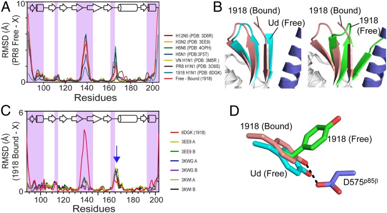
Fig. 5.
Strain-dependent conformational diversity of NS1 proteins. (A) rmsd plots of NS1 proteins from diverse influenza viruses with respect to the structure of PR8 (H1N1, PDB ID code 2GX9). p85β-binding regions are shaded with magenta. X represents individual PDB coordinates. (B) Superimposed structure of p85β-bound 1918 NS1ED and (Left) free Ud NS1ED and (Right) free 1918 NS1ED. (C) rmsd plots of free Ud NS1ED proteins with respect to p85β-bound 1918 NS1ED. Red line: rmsd between free and bound 1918 NS1ED. Blue arrow: residues 164 to 171. (D) Conformations of Y89 in p85β-bound (brown), free (green) 1918 NS1ED, and free Ud NS1ED (cyan).

