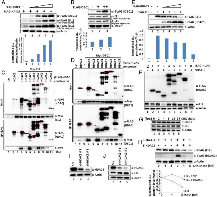
Fig. 2.
DBC1 and HDAC3 act in opposition to regulate ELL stability within mammalian cells. (A and B) Western blot analysis showing the effect of overexpression of DBC1 on (A) ectopic and (B) endogenous expression of ELL in 293T cells (Upper) and quantitation of ELL protein relative to Actin (Lower). (C and D) Co-IP and Western blot analysis showing interactions of HDAC1-8 with (C) ELL and (D) DBC1 in 293T cells. Because of the unavailability of human HDAC2, murine HDAC2 was used in this assay. Red filled circles indicate target protein bands, and others are either degradation or nonspecific proteins. (E) Western blot analysis showing the effect of overexpression of HDAC3 on ectopic expression of ELL in 293T cells (Upper) and quantitation of ELL relative to Actin (Lower). (F) Western blot analysis showing effect of overexpression of HDAC1-8 on ectopic expression of ELL in 293T cells. (G) CHX chase assay showing degradation kinetics of endogenous DBC1 and ELL at different time points after addition of CHX in 293T cells. (H) CHX chase assay showing degradation kinetics of ectopically expressed ELL in presence or absence of ectopically expressed HDAC3 at different time points after addition of CHX in 293T cells (Upper) and quantitation of ELL degradation over time (Lower). (I and J) Western blotting analysis showing (I) the stable knockdown of HDAC3 and (J) its effect on expression of endogenous ELL by using two different shRNAs in 293T cells.

