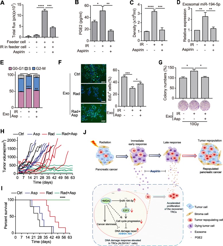
Fig. 6.
Aspirin suppresses tumor repopulation after radiotherapy via impairing the secretions from dying tumor cells. a Bioluminescence intensity of reporter cells that were cultured alone or co-cultured with feeder cells and treated with or without aspirin as indicated. b ELISA detecting the concentration of PGE2 in the supernatant of SW1990 cells that were treated with 10Gy irradiation or irradiation plus aspirin. c Density of exosomes released from SW1990 cells that were treated as in Fig. 6b. d qPCR detection of miR-194-5p in exosomes isolated from SW1990 cells treated as in Fig. 6b. e-f Cell cycle assay (e), representative images (left) and quantifications (right) of EdU incorporation (f) in SW1990 cells treated with exosomes from the indicated cells. Scale bar: 100 μm. g Relative colony numbers (top) and representative images (down) of SW1990 cells treated with exosomes from the indicated cells and subjected to 10Gy radiation. h-i Tumor growth curve (h) and survival rate (i) of PDX tumor-bearing mice that were untreated, or treated with aspirin, 10Gy radiation or radiation plus aspirin. n = 5~6 for each group. j Schematic diagram showing that aspirin disrupts dying tumor cell-orchestrated tumor repopulation cascades. Data are presented as mean with SD of at least three independent experiments; *p < 0.05; **p < 0.01; ***p < 0.001; ****p < 0.0001 from unpaired Student’s t test, expect in Fig. 6i, which was calculated by Log-rank (Mantel-Cox) test

