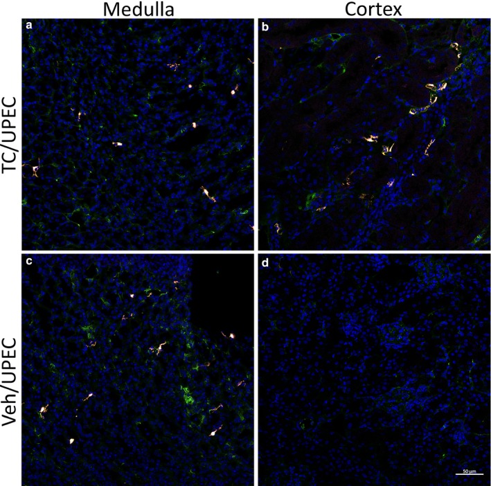
FIGURE 8.

Activated myofibroblasts are localized to the renal cortex only in TC‐treated mice. Immunofluorescence imaging of 8‐µm sections of fixed, frozen kidneys from vehicle‐ or TC‐treated Gli1‐tdTomato mice 28 dpi shows that activated myofibroblasts (αSMA+ (violet), Gli1+ (red), and PDGFRβ+ (green)) and MSC‐like cells (PDGFRβ+ alone) are identified in the medulla in mice receiving either TC (a) or vehicle (c), whereas activated myofibroblasts are observed in the renal cortex only in TC‐treated mice (b), and not in vehicle‐treated mice (d). Scale bar represents 50 µm
