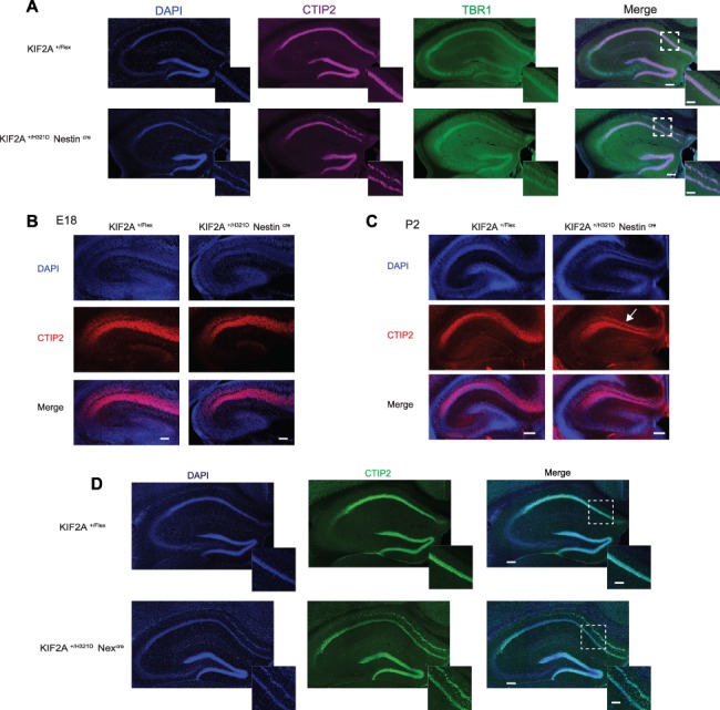
Figure 3.

KIF2A+/H321D mice present with hippocampal heterotopia. (A) Coronal sections of the hippocampus in adult KIF2A+/H321DNestinCre and control mice, stained with CTIP2 (magenta), TBR1 (green) and counterstained with DAPI (blue), scale bar 0.2 cm. Insets delimited with a dotted square show higher magnification of the CA1 region, scale bar 0.1 mm. (B, C) Coronal sections of the hippocampus in E18 KIF2A+/H321DNestinCre and control embryos (B, scale bar 0.1 cm) and P2 pups (C, scale bar 0.2 cm), stained with CTIP2 (red) and counterstained with DAPI (blue). (D) Immunofluorescence staining of CTIP2 (green) and DAPI (blue) on hippocampal slices from adult KIF2A+/H321D Nexcre mice. Scale bar 0.2 cm. Insets delimited with a dotted square show higher magnification of the CA1 region, scale bar 0.1 cm.
