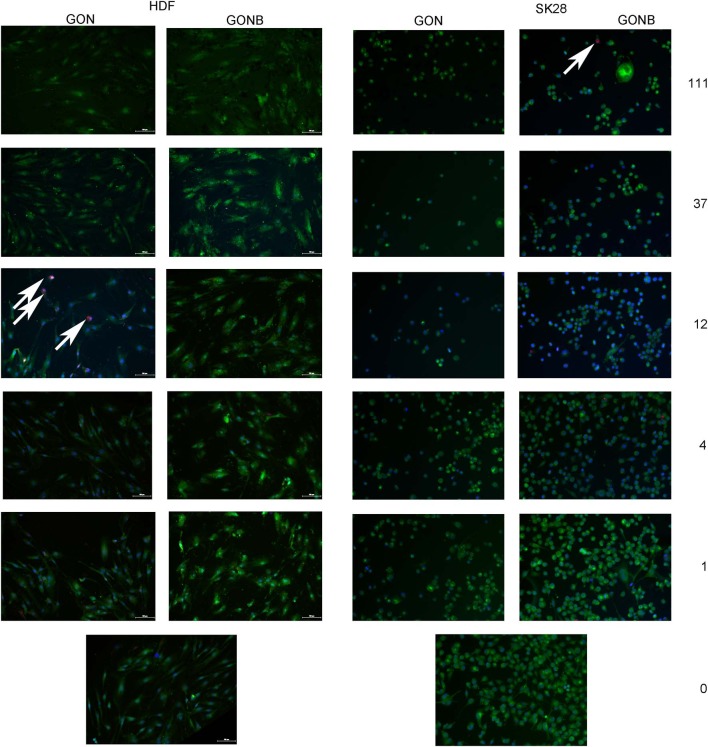
Figure 6.
Representative fields of view obtained by TissueFAXS scanning of GON and GONB solutions treated HDF or SK28 cells, evidencing superior biocompatibility of BSA functionalized GON (GONB) when compared to simple GON. The white arrows indicate the EthD-1 stained dead cells nuclei. Viable cells stained with calcein are visible in green. Scale bar = 100 μm.

