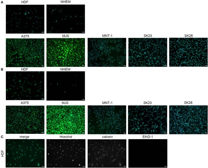
Figure 7.
Immunofluorescence microscopy images obtained by scanning GON (A) and GONB (B) thin films obtained by MAPLE upon culture with normal skin cells and melanoma cells. The images revealed high biocompatibility for both GON and GONB thin films on control and human melanoma skin cells grown after 72 h. Viable cells are shown in green due to calcein staining. Dead cells were not detected. Merged images were obtained by overlapping the TissueFAXS captured fields of view on each fluorescence channel for detection of Hoechst, calcein, and EthD-1 (C). Images correspond to HDFs grown on standard cover slip borosilicate glass. Scale bar = 50 μm.

