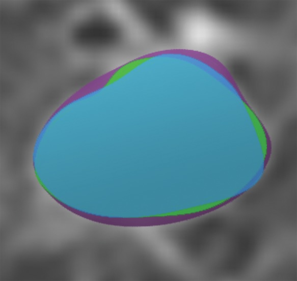
Figure 4c:

Axial images show medical image segmentations performed by experts. (a) CT examination of patient with lung nodule. (b) Nodule is independently and blindly segmented by three medical experts with free open-source software package (Horos, version 3.3.5; Nimble d/b/a Purview, Annapolis, Md). (c) Magnified image of segmentations. There are differences between segmentations; however, these differences are small and not clinically relevant.
