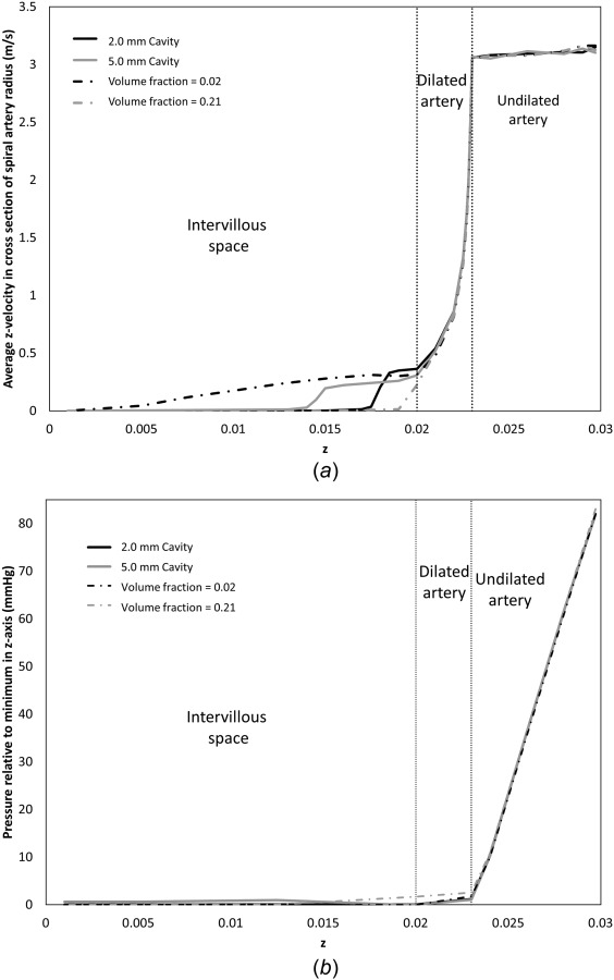
Fig. 6.

Model predictions of (a) average blood flow velocity through the SA and into the placental tissue in a region of the same diameter as the opening of the SA, and (b) average blood pressure in a spiral artery cross section relative to minimum pressure along the z-axis at 25 weeks gestation. Results shown represent a placentome with a 2 mm central cavity (average ψ = 0.21), a 5 mm central cavity (average ψ = 0.21), no central cavity (average ψ = 0.21), and a homogenously reduced tissue volume fraction (ψ = 0.02). All simulations have the same fixed inlet velocity. Visible jet length is sensitive to the structure of villous tissue distal to the SA opening.
