Fig. 3.

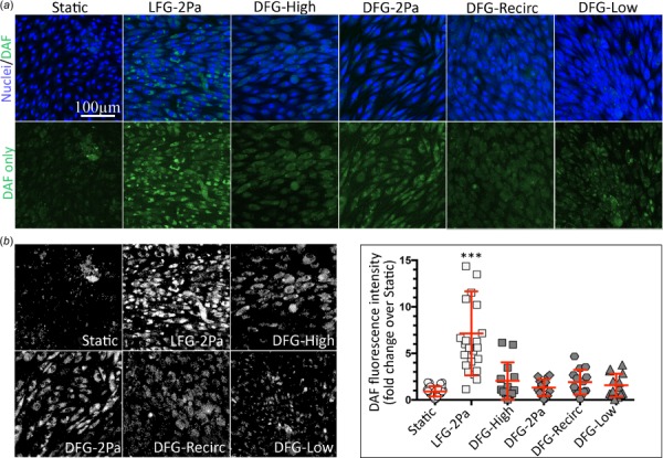
NO was elevated in BAEC after 36 h in the laminar but not disturbed flow parallel plate flow chamber. (a, top) BAEC confocal microscopy images (20×) in varied flow regimes for 36 h showing NO (DAF-FM, green) and nuclei (bisbenzimide, blue). (a, bottom) NO only (DAF-FM, green) confocal microscopy images (20×). (b, left) DAF signal after thresholding to remove noise. (b, right) Total DAF-FM fluorescence intensity quantification for BAEC exposed to static or flow conditions for 36 h (122 images among 18 samples, across 4 independent experiments). *** indicates p < 0.0001 (LFG compared to all other conditions).
