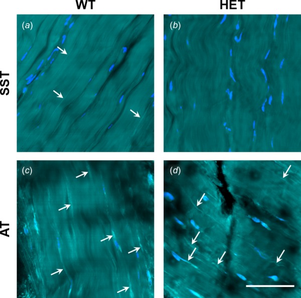
Fig. 3.

Distribution of SRB-stained elastic fibers (bright cyan; white arrows), tenocytes (blue), and collagen (cyan) in WT and HET SSTs and ATs. Few or no fibers were visible in SSTs (a,b). Stained fibers are visible in WT and HET ATs (c,d), where they conformed to collagen orientation and were often localized near tenocytes. No differences were evident between genotypes. Scale bar = 50 μm. Refer to online version for color figure.
