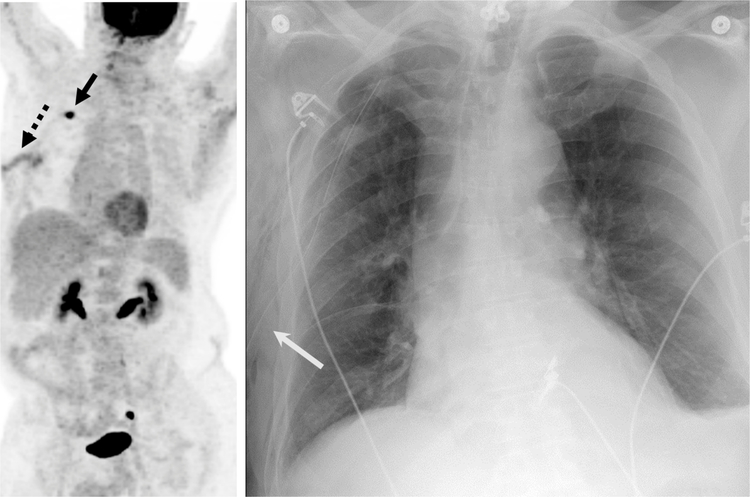
Fig. 12.
67-year-old man with a Recent Diagnosis of Lung Carcinoma.
A, Maximum intensity projection image from an 18F-FDG PET/CT demonstrates a hypermetabolic right upper lobe mass (solid black arrow) with an additional linear focus of uptake in the subcutaneous tissues of the right chest wall (dashed arrow) that was inflammatory due to a right thoracotomy tube placed due to a post-biopsy pneumothorax.
B, Frontal chest radiograph demonstrates the location of the right thoracotomy tube (solid white arrow) confirming the inflammatory etiology of the FDG uptake.

