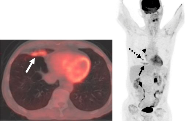
Fig. 3.
Postobstructive 18F-FDG-avid Airspace Disease in a Patient with Lung Malignancy.
A, B Axial fused PET/CT and maximum intensity projection image demonstrates focal uptake in right middle lobe airspace disease (solid arrows) in a patient with a hypermetabolic central right lung nodule (dashed arrow) and an adjacent right hilar lymph node (arrowhead).

