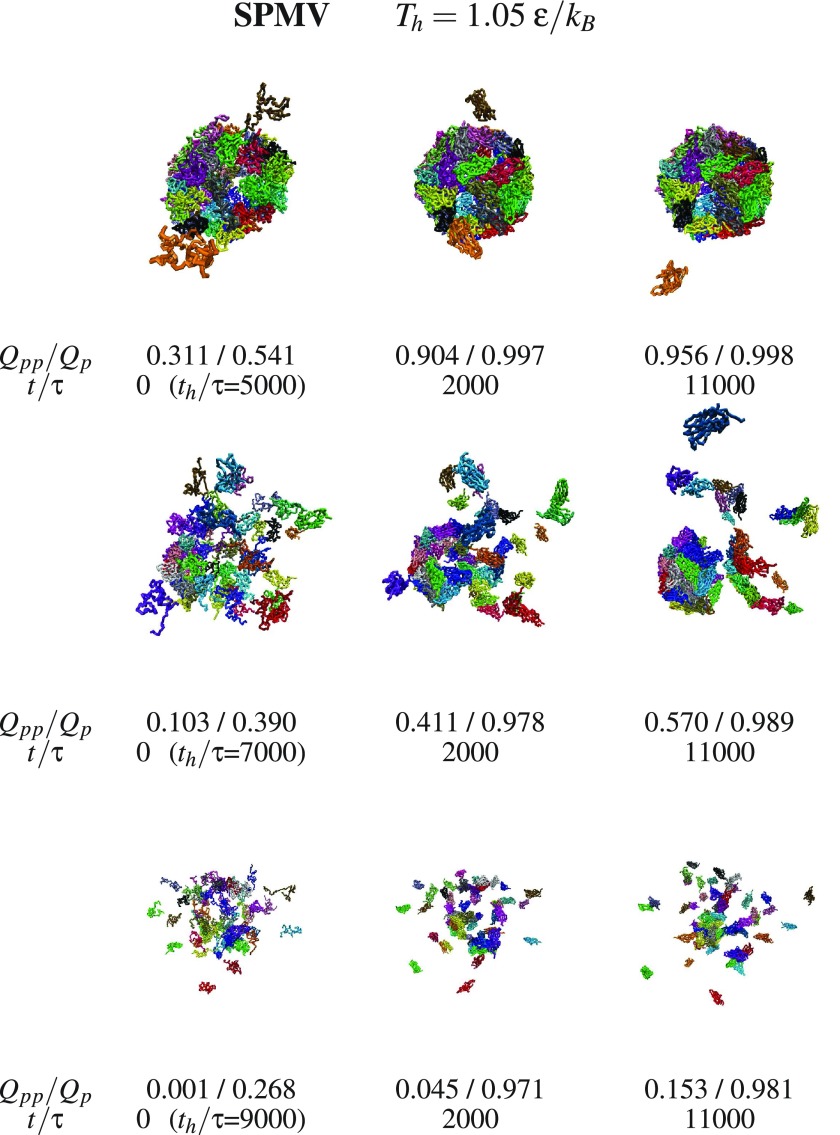
Figure 9.
Examples of the SPMV capsid assembly after thermal denaturation at the temperature indicted at the top. Each horizontal triplet of panels shows snapshots appearing after evolving from the leftmost structure. This starting structure has been obtained at Th applied for time th written underneath in the brackets. The values of Qpp and Qp are indicated. The colors of the proteins are arbitrary.

