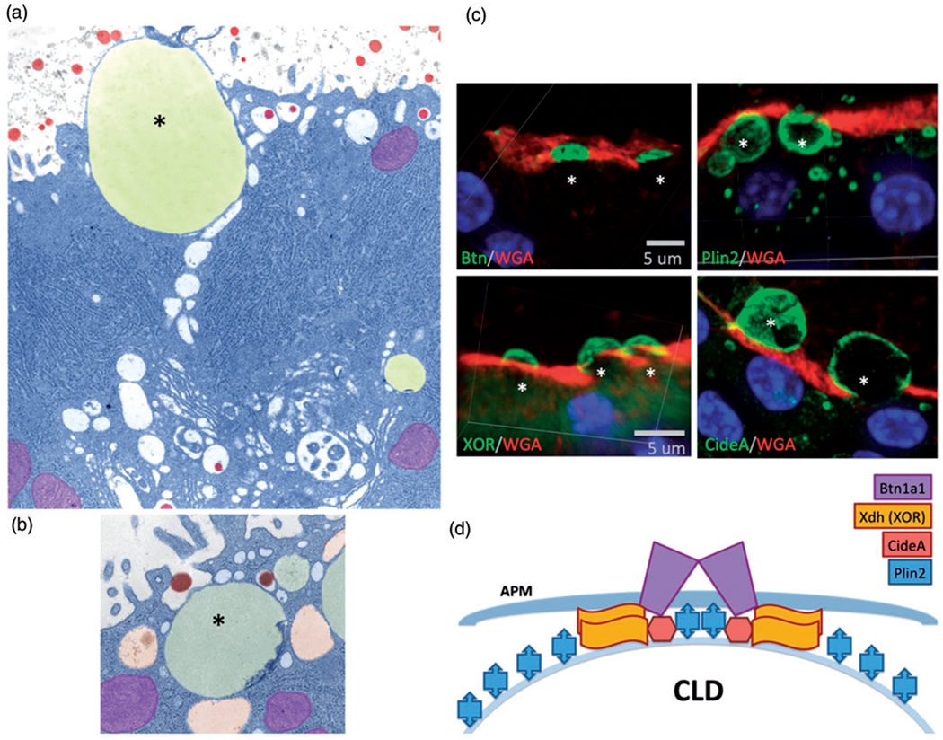
Figure 2.

Cytoplasmic lipid droplet interaction with the apical plasma membrane. (a) Colorized electron micrograph showing a lipid droplet (yellow, asterisk) docked at the apical plasma membrane. Mitochondria are colored purple and casein micelles are colored red. (b) Colorized electron micrograph showing a lipid droplet (asterisk) surrounded by secretory vesicles (orange), and some containing casein micelles (red). (c) Immunofluorescence labeling of proteins in the docking complex that tethers the cytoplasmic lipid droplet to the membrane. BTN, PLIN2, XOR, and CIDEA are shown in green. The membrane is stained with WGA (red), and the nuclei are stained with DAPI (blue). (d) Diagram of the docking complex: BTN is a transmembrane protein (purple), and XDH/XOR is a cytoplasmic protein which binds directly to BTN. CIDEA is a lipid droplet-associated protein that concentrates in the dock, and PLIN2 is a lipid droplet coat protein which becomes covalently cross-linked to BTN and XOR. Stoichiometry was determined by proteomic analysis. WGA = wheat germ agglutinin; CLD = cytoplasmic lipid droplet; XOR = xanthine oxidoreductase; APM = apical plasma membrane.
