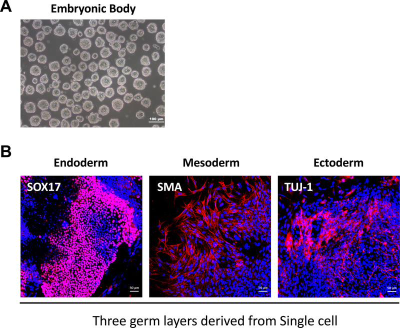
Figure 2: In vitro differentiation of adapted single-cell type hESCs into three germ layers.
(A) Representative phase images of embryonic bodies (EB) derived from single-cell type of hESCs. Scale bar = 100 μm. (B) Immunofluorescent images of differentiated hESCs analyzed for the expression of the three different germ layer markers: SOX17 (endoderm), SMA (mesoderm), and Tuj-1 (ectoderm). Nuclei were stained with DAPI. Scale bar = 50 μm.

