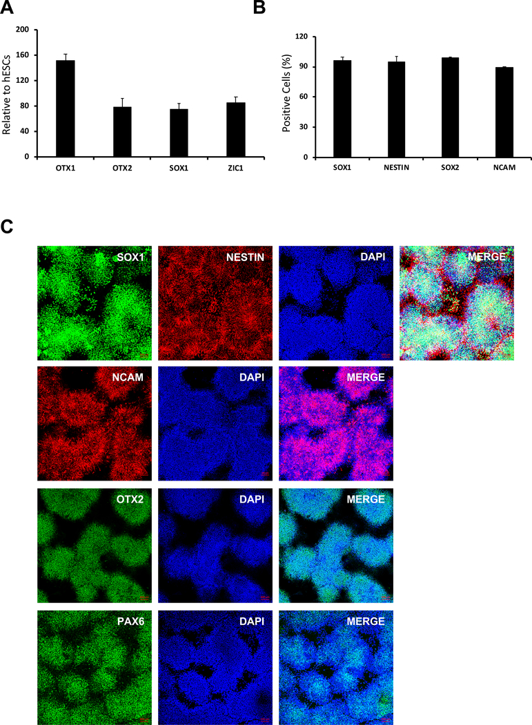
Figure 5: Expression of NPC markers.
(A) After 7 days of neural differentiation, expression of NPC marker genes (i.e., OTX1, OTX2, SOX1, and ZIC1) was analyzed by QRT-PCR. Values were normalized to GAPDH and calculated relative to the values of hESCs (p < 0.05). (B) The percentage of SOX1-, NESTIN-, SOX2-, and NCAM-positive cells was determined by flow cytometry at day 7 of NPC differentiation. (C) At day 7 NPC differentiation, cells were stained with antibodies against the neural markers SOX1, NESTIN, NCAM, OTX2, and PAX6. Nuclei were stained with DAPI. Scale bar = 100 μm.

