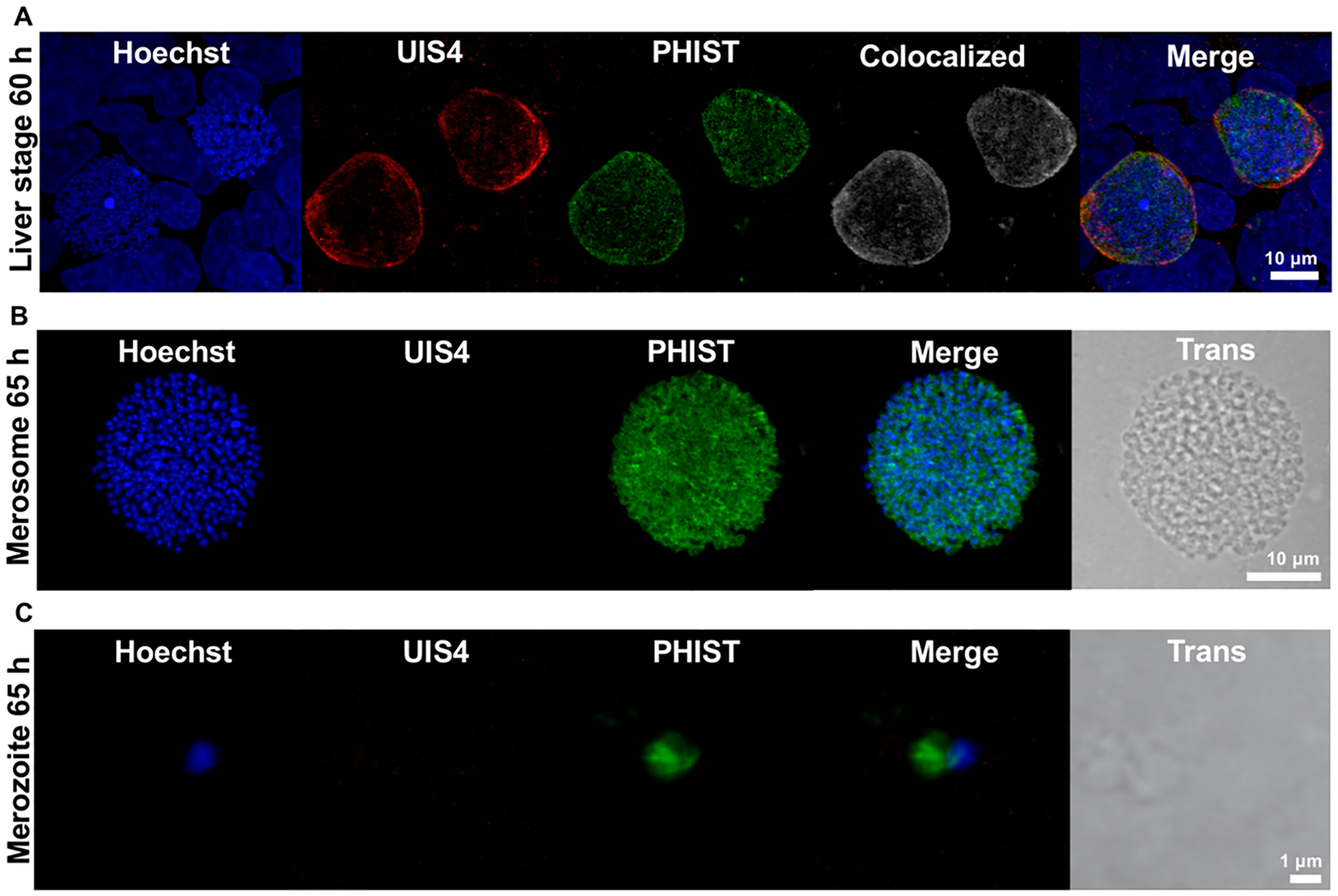
Figure 4.

Localization of the PHIST protein in late liver stage parasites and merosomes. (A) Immunofluorescence staining of late liver stage parasites at 60 h postinfection indicates the PHIST protein (PBANKA_1145400) partially colocalizes with the parasitophorous vacuole marker UIS4. Colocalized pixels in white. DNA stained with Hoechst. (B,C) Immunofluorescence staining of merosomes and free merozoites at 65 h postinfection demonstrates the PHIST protein associates with individual liver stage merozoites. No UIS4 staining was detected in the mature merosome or free hepatic merozoite shown.
