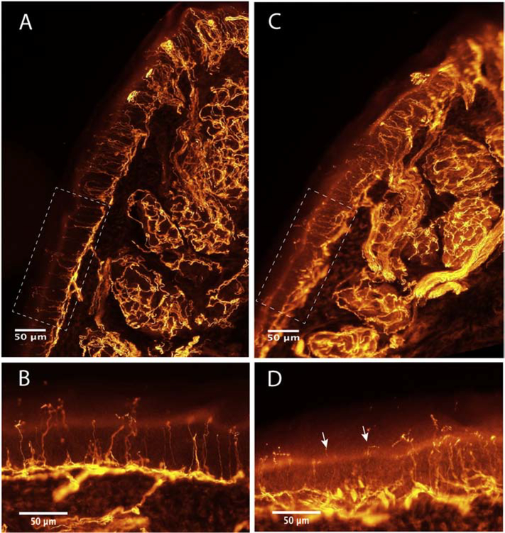
Figure 3.
Immunohistochemical images of the plantar pad of a control mouse (A, B) and a mouse treated with cisplatin (C, D), labeled against PGP9.5. The images show densely innervated sweat glands in the dermis, the subepidermal nerve plexus underlying the epidermis, the Meissner corpuscles at the papillae of the pad tip (top part of the pad), and the numerous intraepidermal nerve fibers (IENF) crossing the epidermis. Boxes in A,C are the area of epidermis magnified in B,D. Note the reduction in number of IENF and the degenerating appearance of some IENF (arrows) in the skin of the cisplatin-treated mouse.

