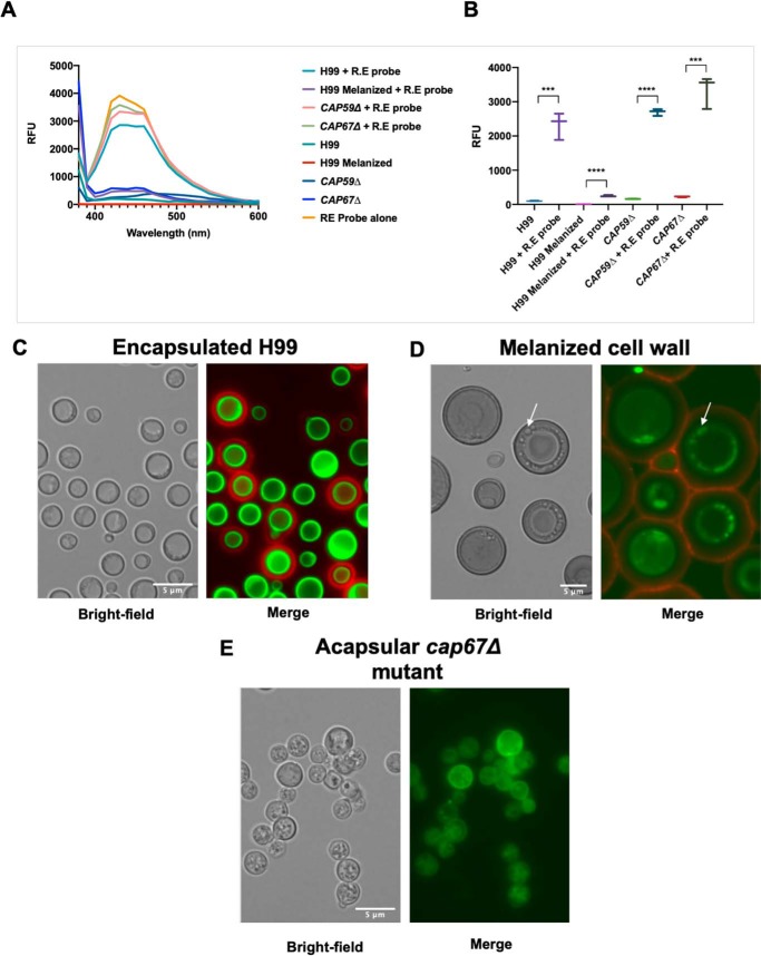
Figure 2.
Incubation of hydroxylamine-armed fluorescent probe with C. neoformans. A, fluorescence spectrum of C. neoformans cells incubated with and without hydroxylamine-armed probe (R.E probe). Those that have been incubated with the R.E probe have increased emission maxima at ∼453 nm (excitation 360 nm) compared with encapsulated H99, CAP59Δ, or CAP67Δ with the exception of C. neoformans with encapsulated H99 with a melanized cell wall. Melanization of the cell wall is likely quenching the florescence of the probe. B, RFU of C. neoformans cells incubated with the R.E probe are significantly higher at emission maxima (440 nm) of the probe (excitation 360 nm). Error bars represent 95% confidence interval. Statistical significance was determined using unpaired t test (***, p ≤ 0.001; ****, p ≤ 0.0001). Experiments were performed in triplicate. C, encapsulated H99: the capsule shown by anti-GXM Mab 18B7-AF594 (red) displays no reactivity to the R.E probe 6 (green) and is localized at the cell wall–membrane interface. D, melanized C. neoformans cells display the same localization of the R.E probe. The R.E probe appears intracellularly possible in vesicular bodies and displays no reactivity toward the capsule, despite the melanization of the cell wall. E, acapsular mutant C. neoformans cap67Δ incubated with the R.E probe shows bright fluorescence intensity coming from the cytoplasm and the cell wall–membrane interface. Labeling of cytoplasm occurs in spherical vesicle-like structures in acapsular cap67Δ and melanized H99 cells. Scale, 5 μm.

