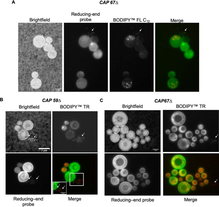
Figure 5.
Secretion of vesicular bodies in CAP59Δ and CAP67Δ mutants. A, incubation of CAP67Δ with BODIPY FL C12 (lipophilic dye, red) and the R.E probe shows vesicle secretion (arrows). B, CAP59Δ cells incubated with BODIPY TR (lipophilic dye, red) also show signs of secretion of glycan-containing vesicles (inset and arrows). C, CAP67Δ cells incubated with the BODIPY TR and R.E probe show signs of colocalization; however, BODIPY TR also stains nonglycan-containing vesicles (arrows) and shows signs of secretion of nonglycan-containing vesicular bodies (arrow). Scale denoted in bright field: A, 5 μm; B, 7 μm (inset, 5 μm); and C, 5 μm.

