Figure 3.

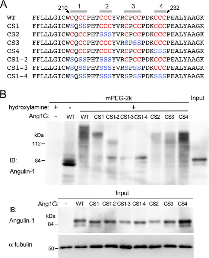
Determination of palmitoylated cysteine residues in angulin-1. A, amino acid sequences of the juxtamembrane regions in mouse angulin-1 (WT) and its mutants with serine substitution of the cytoplasmic cysteine residues (CS1, CS2, CS3, CS4, CS1–2, CS1–3, and CS1–4). The cysteine and serine residues within amino acids 210–232 are shown in red and blue letters, respectively. B, the top panel shows palmitoylation of GFP-tagged WT angulin-1 and its cysteine-to-serine–substituted mutants detected by the APEGS method. Lysates of Ang1KO cells stably expressing Ang1G (WT) or mutants were processed for the APEGS method. Mobility shifts of the bands to higher molecular weights indicate protein palmitoylation. The bottom panel shows immunoblotting of lysates from Ang1KO cells (−) and cells expressing Ang1G (WT) or cysteine-to-serine–substituted mutants before the procedure for the APEGS method with an anti-angulin-1 antibody and anti-α-tubulin antibody. IB, immunoblotting.
