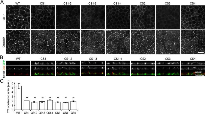
Figure 4.
Role of palmitoylated cysteine residues in the localization of angulin-1 at TCs. A, immunofluorescence staining of Ang1KO cells stably expressing Ang1G (WT) or cysteine-to-serine–substituted mutants with an anti-occludin antibody. Ang1G or the mutants in the same field were also visualized by fluorescence of GFP. Scale bar, 20 μm. B, Z-stack sections of immunofluorescence staining of Ang1KO cells stably expressing Ang1G (WT) or cysteine-to-serine–substituted mutants. The GFP signal (green) from each of the cysteine-to-serine–substituted mutants is connected to the occludin signal (red) and extends in the basal direction, indicating that these mutants are localized along the lateral plasma membrane. Scale bar, 5 μm. C, quantitation of the TC enrichment of Ang1G (WT) or cysteine-to-serine–substituted mutants. The graph represents mean ± S.D. (error bars) (n = 2–3 each). **, p < 0.005, compared by t test. a.u., arbitary units.

