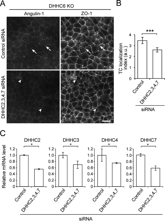
Figure 5.

Role of DHHCs in the localization angulin-1 at TCs. A, immunofluorescence staining of DHHC6 KO cells treated with control siRNA or the mixture of DHHC2, -3, -4, -7 siRNAs with anti-angulin-1 and anti-ZO-1 antibodies. Compared with clear localization of angulin-1 at TCs in control siRNA-treated cells (arrows), the angulin-1 signal at TCs in DHHC2, -3, -4, -7 siRNA–treated cells was faint overall, although part of the TCs showed significant localization of angulin-1 (arrowheads). Scale bar, 20 μm. B, quantitation of the TC enrichment of angulin-1 in control and DHHC2, -3, -4, and -7 siRNA–treated DHHC6 KO cells. The graph represents mean ± S.D. (error bars) (n = 6 each). a.u., arbitary units. C, relative mRNA expression level of DHHC2, -3, -4, or -7 in DHHC6 KO cells treated with control siRNA or siRNAs for DHHC2, -3, -4, and -7 was measured by qPCR. Ribosomal protein L5 expression was used for normalization. The graph represents mean ± S.D. (n = 3 each). *, p < 0.05; ***, p < 0.0005, compared by t test.
