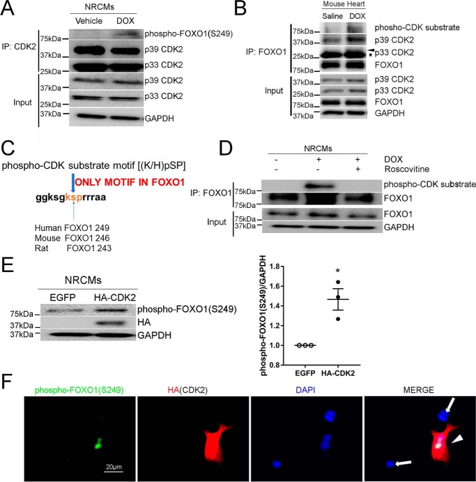
Figure 2.
Stimulation with DOX enhanced FOXO1 phosphorylation by CDK2. A, NRCMs were treated with DOX (1 μm) or vehicle for 4 h. Protein lysates were immunoprecipitated (IP) with anti-CDK2 antibody followed by Western blotting with the indicated antibodies. B, adult C57BL/6 mice were injected with DOX (5 mg/kg, i.p.) or saline and euthanized at 24 h. Protein lysates were immunoprecipitated with anti-FOXO1 antibody followed by Western blotting. Asterisk, IgG light chain. C, protein sequence analysis revealed that only one phospho-CDK substrate motif, (K/H)pSP, exists in FOXO1. D, NRCMs were pretreated with the CDK inhibitor roscovitine (50 μm) for 16 h prior to incubation with DOX (1 μm) for 4 h. Protein lysates were immunoprecipitated with anti-FOXO1 antibody followed by Western blotting. E, NRCMs were transfected with EGFP or HA-CDK2, and protein levels were measured by Western blotting. Two-tailed Student's t test. *, p < 0.05 versus EGFP. F, NRCMs transfected with HA-CDK2 were subjected to immunofluorescence staining for phospho-FOXO1 (Ser-249, green), HA tag (red), and nuclei (DAPI, blue). An intense phospho-FOXO1 (Ser-249) signal was observed in HA-positive cells (arrowhead) but not in HA-negative cells (arrows). Scale bar = 20 μm.

