. 2020 Mar 30;9:e53387. doi: 10.7554/eLife.53387

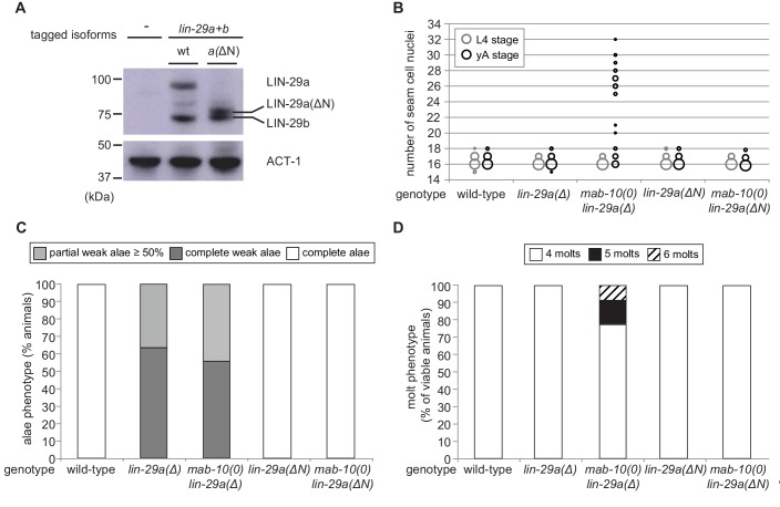
Figure 6. The LIN-29a-specific domain is dispensable for the execution of the J/A transition.

(A) Western blot of endogenous C-terminally GFP::3xFLAG-tagged LIN-29a and LIN-29b proteins in the lin-29a(ΔN) background (HW2408) using an anti-FLAG antibody. Actin-1 is used as loading control. (B) Seam cell number quantification in L4 larval stage and young adult (yA) animals of the indicated genetic backgrounds (n > 25 for L4, n > 25 for yA worms per genotype). The data for lin-29a(Δ) and mab-10(0) lin-29a(Δ) is re-plotted from Figure 2 for comparison. (C) Quantification of different alae structures in young adult worms of indicated genotypes (n > 20). The data for lin-29a(Δ) and mab-10(0) lin-29a(Δ) is re-plotted from Figure 3 for comparison. (D) Quantification of the number of molts for animals of indicated genotypes (n > 20). The data for lin-29a(Δ) and mab-10(0) lin-29a(Δ) is re-plotted from Figure 3 for comparison. In (B – D), lin-29(∆N) is lin-29(xe200).

Figure 6—source data 1. Quantification of seam cell numbers, raw data related to Figure 6B.
Figure 6—source data 2. Quantification of alae structures, raw data related to Figure 6C.
Figure 6—source data 3. Quantification of number of molts, raw data related to Figure 6D.

Figure 6.

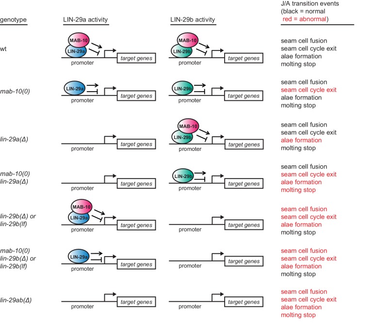
Figure 6—figure supplement 1. Summary of the J/A transition phenotypes seen for different permutations of lin-29a, lin-29b, and mab-10 mutations.

Figure 6—figure supplement 1.

Note that extra seam cell divisions in mab-10(0) mutant animals occur only in older adults. Some older mab-10 mutant adults may also undergo extra molts (Harris and Horvitz, 2011), although we did not observe this.
Figure 6—figure supplement 2. Characterization of lin-29a(∆N) expression and function.

Figure 6—figure supplement 2.

(A) Schematic representation of the lin-29a(ΔN) deletion. (B) Micrographs of late L4-stage animals of indicated genotypes expressing scm::gfp (green, marking seam cells) and ajm-1::mCherry (red, marking hypodermal cell junctions). In both lin-29a(xe200[lin-29a(ΔN)]) and mab-10(0) lin-29a(xe200) animals, fusion occurs normally. Scale bar: 50 μm. (C) Western blot of C-terminally GFP::3xFLAG-tagged endogenous LIN-29a and LIN-29b proteins in a wild-type and the lin-29a(ΔN) background (HW2408), respectively, using anti-FLAG antibody. Animals were grown for 20 hr at 25°C to the L3 stage on mock RNAi and lin-41 RNAi bacteria, respectively. Both LIN-29a and LIN-29a(∆N) accumulate upon knock-down of lin-41. Actin-1 is used as a loading control. (D) Confocal images of endogenously tagged LIN-29 protein isoforms in the wild type and lin-29a(ΔN) background (HW2408) in the epidermis of animals at the indicated developmental stages. Animals were staged by examination of gonad development. Arrows indicate seam cell, arrowheads hyp7 nuclei. Scale bars: 10 μm.
Figure 6—figure supplement 2—source data 1. Quantification of unfused seam cell junctions, raw data related to Figure 6—figure supplement 2B.