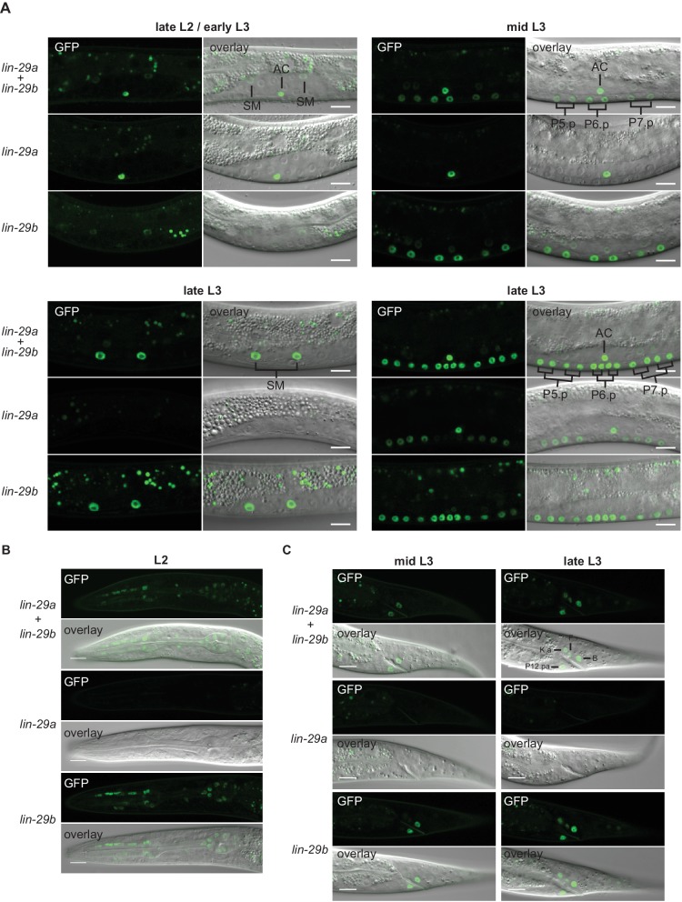
(A) Confocal images of endogenously tagged LIN-29 protein isoforms in the region of the vulva and the uterus at the indicated developmental stages. At the L2-to-L3 molt (A), lin-29a is expressed in the anchor cell (AC), while lin-29b is weakly expressed in the sex myoblasts (SMs). In mid-L3 stage worms, the six daughters of the VPCs P5.p-P7.p express lin-29b. At the late L3 stage, lin-29b is strongly expressed in the sex myoblast (SM) daughters and all 12 granddaughters of the VPCs P5.p-P7.p, while LIN-29a specifically accumulates in the granddaughters of P5.p and P7.p, but not in those of P6.p. Scale bars: 10 μm. (B) Confocal images of endogenously tagged LIN-29 protein isoforms in the pharynx of L2 stage animals at the indicated developmental stages. lin-29b is expressed in the pharynx throughout larval and adult development. Scale bars: 10 μm. (C) Confocal images of endogenously tagged LIN-29 protein isoforms in the tail region at the indicated developmental stages. LIN-29b first accumulates in the two rectal cells B and P12.pa, before accumulating in the four additional rectal cells F, K.a, K’ and U (the latter two are not visible in this focal plane). LIN-29a is not detected in these cells. Scale bars: 10 μm.


