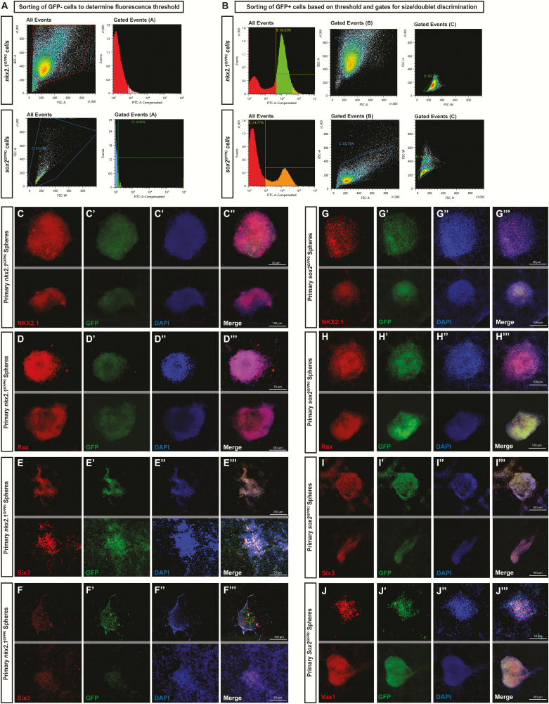
Figure 5.
Validation of hypothalamic identity of FACS-generated neurospheres.
FACS methodology is displayed for sorting GFP- cells to establish a fluorescence threshold (A) and for sorting GFP+ cells to isolate single cells for culturing (B). Neurospheres cultured from tissue dissected from NKX2.1GFPKI transgenic mice express the hypothalamic markers Nkx2.1 (C-C′′′), Rax (D-D′′′), Six3 (E-E′′′), and Vax1 (F-F′′′). Similarly, neurospheres cultured from tissue dissected from Sox2GFPKI also express Nkx2.1 (G-G′′′), Rax (H-H′′′), Six3 (I-I′′′), and Vax1 (J-J′′′). (2 representative fields are presented; grey lines divide different fields of exposure).

