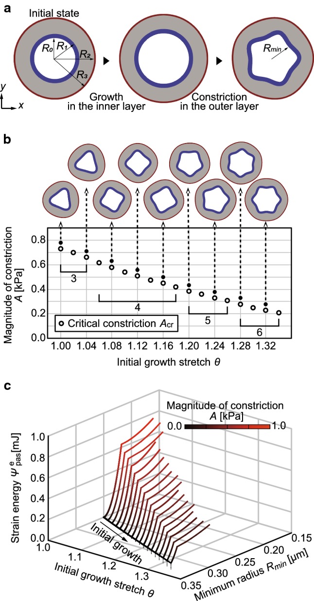
Fig. 4.

Application to luminal folding in the initial stage of intestinal villus formation. a Typical luminal folding process induced by circumferential growth in the inner layer followed by circumferential constriction in the outer layer. The inner layer in blue is the epithelium. The middle layer in gray is the mesenchyme. The outer layer in red is the muscle. b Critical magnitude of constriction , at which the tube folded, as shown in open circles. Snapshots pointed by arrows are the morphologies at each solid circle. c Energy landscape of luminal folding, which shows the relationship between the total strain energy in the inner layer [mJ], minimum radius of the inner layer [μm], and growth stretch . The color map represents the magnitude of constriction A [kPa]
