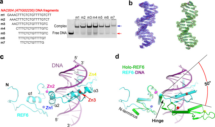
Fig. 3. Structure of the REF6-DNA binary complex.
a REF6 binds to NAC004 dsDNA of different lengths with the same concentration. Positions of free dsDNA and protein-bound DNA are indicated by red and blue arrows, respectively. b The composite simulated-annealing sigma-A-weighted 2mFo − DFc (left) and sigma-A-weighted mFo − DFc (right) electron density maps are shown at 1.5σ and 2.5σ, respectively. c Ribbon representation of the REF6-DNA structure. REF6 and DNA are in blue and purple color, respectively. d Superposition of the holo-REF6 (green) and the REF6-DNA complex (REF6 in cyan and DNA in purple) structures. The coordinates of the RCZ domains from each were aligned. The last two ZnF domains showed a rotation of ~50° towards the dsDNA.

