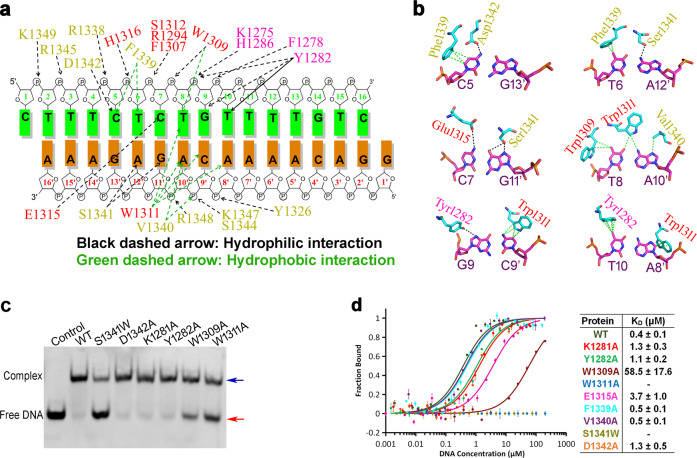
Fig. 4. Interaction of REF6 and dsDNA.
a Schematic representation summarizing the REF6−DNA interactions. The sequence of the dsDNA (NAC004 fragment) used for crystallization is shown with two complementary strands. The residues involved in the interaction with the dsDNA are labeled in magenta, red, and yellow for domains ZnF2, 3, and 4, respectively. The green and black dashed arrows indicate hydrophobic and hydrophilic interactions, respectively. b Close-up view of the interaction between key residues and DNA bases. c EMSA of different REF6 mutants with the dsDNA. d Measurement of the binding affinity of the WT and mutant REF6 with dsDNA by MST. The experiments were repeated for three times.

