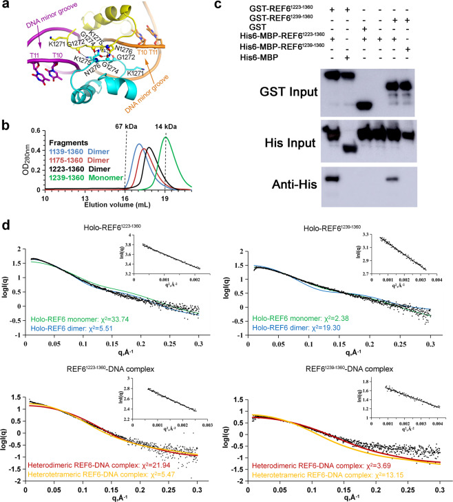
Fig. 6. Self-association of REF6 with or without dsDNA.
a The interface between two REF6 proteins in the complex structure. b Analytical gel filtration profiles of the REF6 fragments on a Superdex 200 10/300 GL column at 5.0 mg mL−1. The fragments of 1139–1360, 1175–1360, 1223–1360 and 1239–1360, all containing the four ZnF domains with different N-terminal extensions, are labeled in blue, red, black, and green, respectively. Elution volumes of the molecular mass standards are marked at the top of the panel. c Various fragments of purified GST-REF6 were used for GST pull-down of purified His-REF6 in the presence of NAC004 DNA fragment. Input and eluted proteins were analyzed by Western blot analysis with the anti-His antibody. d Comparison of SAXS experimental data and calculated scattering profiles. The inset shows the Guinier fits of 5 mg ml−1 holo-REF6 and REF6-DNA complex. Experimental data are represented in black dots. Holo-REF6 monomer (green), holo-REF6 dimer (blue), heterodimeric REF6-DNA complex (red), heterotetrameric REF6-DNA complex (yellow). Note that holo-REF6 dimer and heterotetrameric REF6-DNA complex models are built based on the structures obtained by the symmetric operation.

