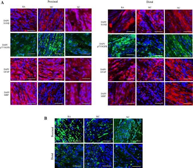
Fig. 4.
IHC analysis (ICC) of the sciatic nerve 12 weeks post-implantation for RA, MC and NC groups (proximal and distal). Nuclei were counter-stained with DAPI (blue). Analysis of the longitudinal section. Magnification, 20×. Scale bar, 100 μm. n = 3. A ICC of the sciatic nerve for protein S100β (red), p75NGFR (green), GFAP (red) and MBP (red). B ICC of the sciatic nerve for protein NF (green)

