Fig. 6.

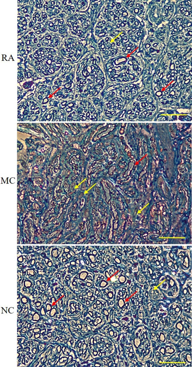
Transverse section of toluidine blue-stained sciatic nerve under a light microscope. Red arrows show myelinated axons, whereas yellow arrows show non-myelinated axons. Magnification, 40×. Scale bar, 200 μm. n = 3

Transverse section of toluidine blue-stained sciatic nerve under a light microscope. Red arrows show myelinated axons, whereas yellow arrows show non-myelinated axons. Magnification, 40×. Scale bar, 200 μm. n = 3