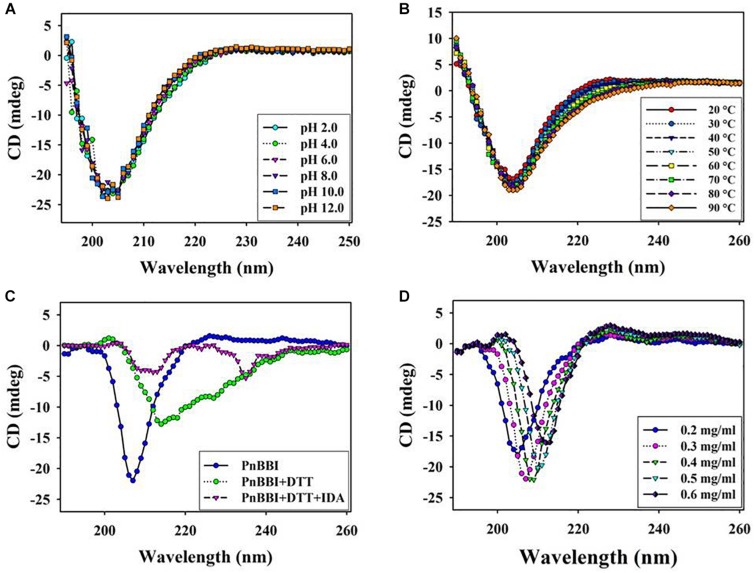
FIGURE 6.
Structural stability of PnBBI. Far-UV (190-260 nm) secondary structure of PnBBI was determined by CD spectroscopy at different (A) pH from 2.0 to 12.0; (B) temperatures from 20 to 90°C; (C) DTT (2 mM) reduction followed by IDA (4 mM) alkylation and (D) increasing concentration (0.2 - 0.6 mg/ml) of PnBBI prepared in 10 mM PBS, pH 7.4 as described in methods. The final spectrum is an average of three scans as described in Section “Circular Dichroism Spectroscopy.”

