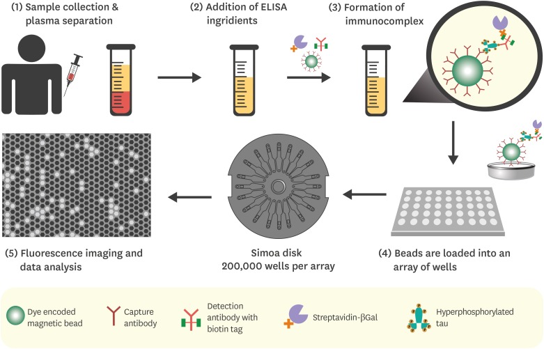
Fig. 3. Quantification of plasma tau using Simoa HD-I analyzer. (1) Blood sample is collected and centrifuged. (2) Paramagnetic microbeads coupled to detection antibodies (supplied along with Simoa Human tau immunoassay kit) are added to the plasma sample, preceding the addition of standard ELISA reagents. (3) If hyperphosphorylated tau protein is present (the target of the immunoassay) in the plasma sample, the formation sandwich immunocomplex will occur. (4) The microbeads are then concentrated by magnetic separation and loaded onto the arrays of femtomolar-wells, each capable of fitting a single immunocomplex. The arrays are located on the Simoa disk composed of 24 arrays. (5) After adding a fluorogenic substrate and sealing the wells using an oil solution, a single-binding event is detected and quantified by the instrument.
ELISA: enzyme-linked immunosorbent assay.

