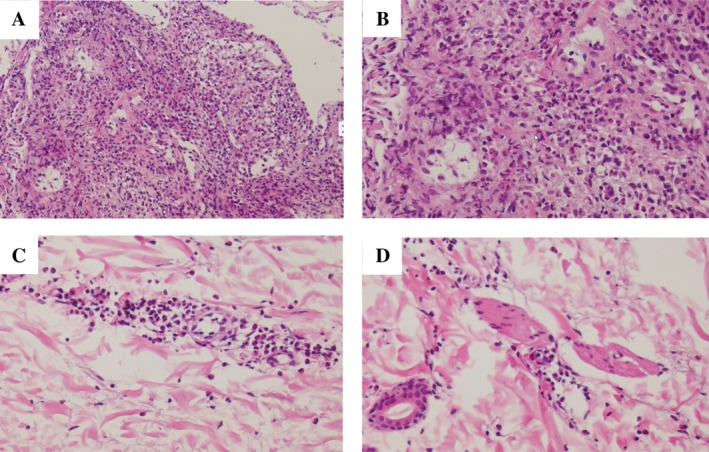
Figure 2.

Pathological findings in a 51‐year‐old woman with eosinophilic granulomatous polyangiitis. (A, B) In the lungs, there are inflammatory granulations with marked eosinophil infiltration (haematoxylin–eosin stain: A, 40×; B, 100×). (C, D) In the skin, there is eosinophilic infiltration and perivascular dermatitis (haematoxylin–eosin stain: C, 100×; D, 100×).
