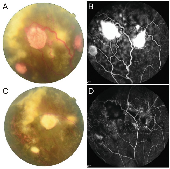
Fig. 1. Clinical outcome of a case 1 patient. (A) Fundus photographs and (B) fluorescein angiography of the right eye of a 29-year-old patient at the initial visit. Three round, orange retinal masses <1.5 disc diameters with dilated feeder vessels and exudations were observed in the peripheral retina. Leakage of fluorescence associated with the lesions was revealed by fluorescein angiography. (C) The size of the masses and exudates decreased after two laser photocoagulation treatments. (D) Leakage around the lesions also decreased after two laser photocoagulation treatments.

