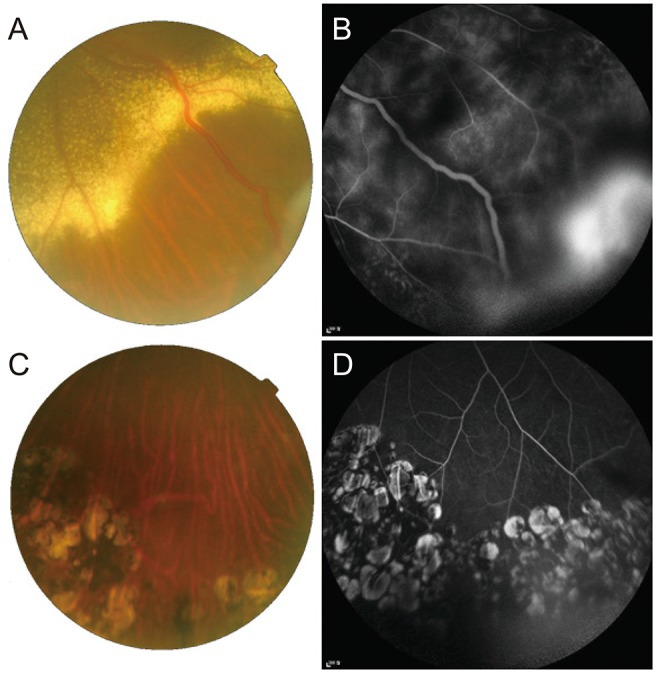
Fig. 3. Clinical outcome of a case 3 patient. (A) Fundus photograph and (B) fluorescein angiography (FA) of the left eye of a 35-year-old patient before cryotherapy. There were exudations with dilated feeder vessels, and (B) FA revealed leakage from the lesion. Seven years after cryotherapy and additional laser photocoagulation treatments, a (C) fundus photograph and (D) FA showed no dilated vessels, exudates, or leakage around the mass.

