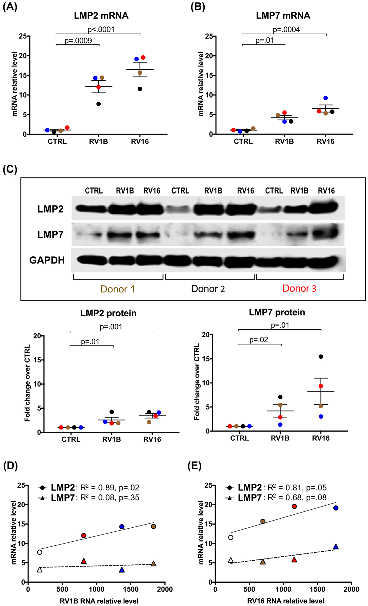
Figure 1. Human tracheobronchial epithelial (HTBE) cells infected with rhinovirus increase immunoproteasome subunit expression.

HTBE cells (n=4 subjects, as represented by different color symbols) grown at ALI were infected with RV1B and RV16 or treated with 0.25% BSA-PBS (control, CTRL) for 24 and 48 h. mRNA levels of IP subunits LMP2 (A) and LMP7 (B) were quantitated by RT-PCR (24 h) and were normalized to GAPDH gene. (C) Representative Western blot images and densitometry of LMP2 and LMP7 proteins at 48 h post RV infection. Correlations between RV1B (D) and RV16 (E) load and the levels of LMP2 and LMP7 mRNA. Data were presented as means ± S.E.M. and analyzed using ANOVA.
