
Fig. 2.
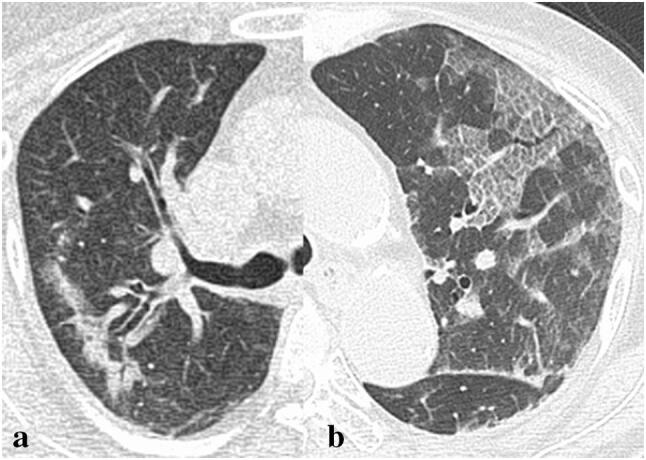
CT images from patients with negative RT-PCR test for COVID-19. a A 66-year-old female patient presenting with persistent fever (day 8). CT shows curved linear consolidation with peripheral distribution as well as bronchial wall thickening. This patient was diagnosed as Moraxella Pneumonia. b A 70-year-old female patients with pancreatic cancer presenting with fever (day 7). CT shows extensive ground glass lesion with crazy paving pattern in left upper lobe. Note these shadows are not strongly in peripheral distribution. The patient was diagnosed as pnuemocystis jirovecii pneumonia