Fig. 1.
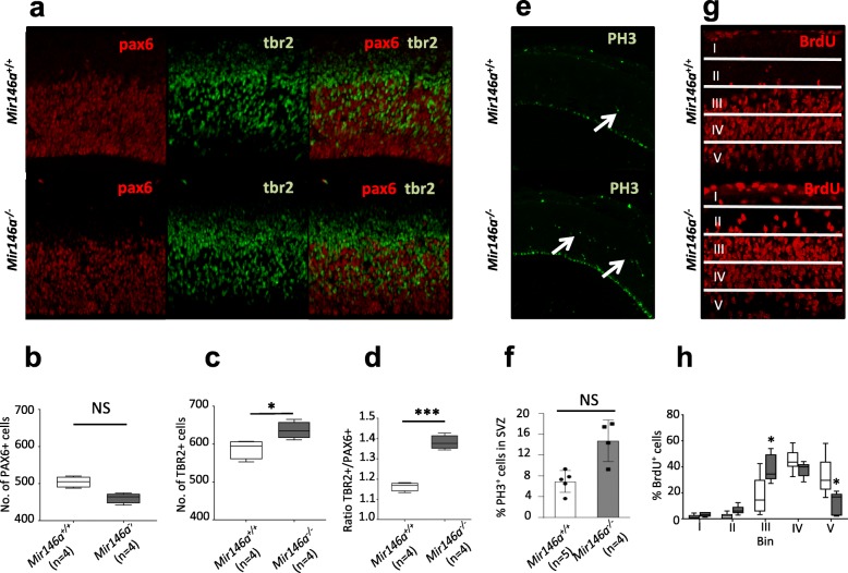
Altered aRG and IP population size in the Mir146−/− brains. a Sagittal sections from Mir146a+/+and Mir146a−/− brains at E14.5 labeled with PAX6, TBR2, and merge. b Boxplot showing the number of PAX6+ cells (±S.D.) counted using 3 images from comparable plane per mouse. c Boxplot showing the quantification of TBR2+ cells (±S.D.) from the same images as b. d Boxplot showing the ratio of TBR2+/PAX6+ cells. e Phospho-histone 3 (PH3) stains dividing cells in the neocortex at E14.5 (white arrows). f Graph showing the average number of PH3+ cells (±S.D.) in the SVZ. *P < 0.05 by Student’s unpaired 2-tailed t test. g BrdU staining in the neocortex at E14.5 after 2h pulse chase; images were divided into five equal bins for counting. h Percentage of BrdU+ cells (±S.D.) per bin (normalized against the total number of labeled cells). NS not significant; *p ≤ 0.05, **p ≤ 0.01, ***p ≤ 0.001 by Mann-Whitney U test

