Fig. 3.
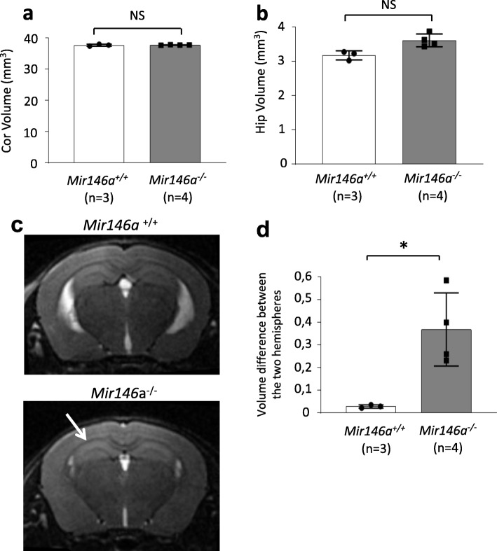
Increased hippocampus asymmetry in the Mir146a−/− brain at P30. Volume (±S.D.) of the hippocampus (a) of the mutant and WT mice as measured by brain MRI at P30. The surface area of each region was measured from MRI images at 100 μM apart (5 images for the hippocampus and 15 for the cortex) and reconstructed using Image J to obtain final volume. b Brain MRI showing coronal sections. White arrows point towards the hippocampi; the larger white arrow point towards the enlarged hippocampus in the mutant. c Graph showing the volumes difference between two hippocampal hemispheres in Mir146a+/+ and Mir146a−/− brains. *p < 0.05 by Student’s unpaired 2-tailed t test. On average, one hippocampus side is 15% larger than the other in Mir146a−/− brains with random distribution (2 right > left and 2 left > right)

