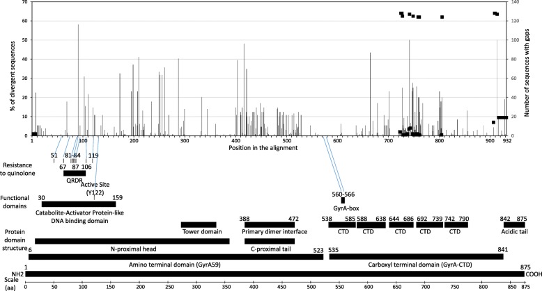
Fig. 2.
Amino acid divergence among the GyrA protein based on 129 unique sequences. The positions within the 932 amino acid long alignment are indicated on the X axis. The percentage of sequences featuring a particular divergent position is indicated on the Y axis (left side). For example, 58% of the sequences feature a mutation at position 91. The divergence plot was generated from the amino acid difference table shown in Additional Table 2. The alignment features 10 gaps and the number of sequences featuring gaps is indicated on the right axis and shown as a black square. A map of E.coli GyrA protein featuring structural and functional domains is shown at the bottom for comparison. The map was generated based on the following references [10, 12, 13]. The GyrA protein sequence of E.coli and N.meningitidis reference strain 053442 (GB-ID CP000381) were compared (Additional Table 4). The known CIP-resistant site in E.coli are shown and corresponding positions in N.meningitidis are indicated with a blue line. It is worth noting that among the 8 resistant sites reported in E.coli, only positions 83 and 87 are divergent in N.meningitidis (sites 91 and 95)

