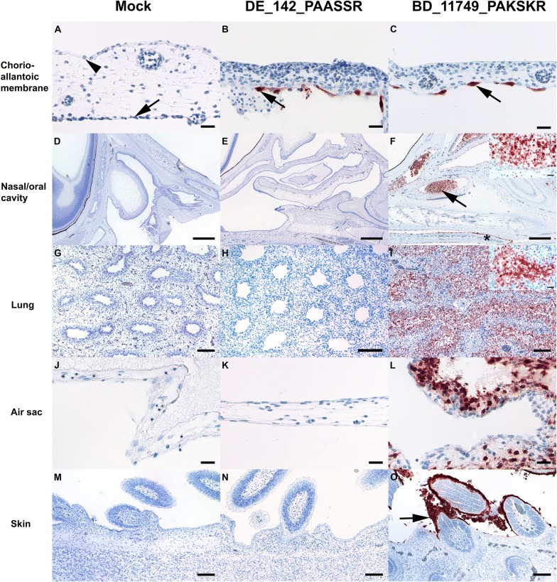
Figure 3.
Immunohistological findings in mock (n = 2), DE_142-PAASSR (n = 2) and BD_11749_PAKSKR (n = 3) avian influenza virus (AIV) infected 14 days old chorio-allantoic membranes and chicken embryos. A Normal cuboidal epithelium (arrowhead) of the chorion and flattened epithelium (arrow) of the allantois. B, C Intense matrixprotein immunoreactivity confined to allantoic epithelium (arrow). D, E Nasal cavity and a section of the eye in the left upper corner. F Necrotizing rhinitis with marked luminal accumulation of matrixprotein positive debris in the rostral (arrowhead) and caudal nasal chamber as well as immunoreactive squamous epithelium of the tongue (star); Inset: The lumen of the nasal cavity is filled by epithelial debris and remaining respiratory epithelium is either flattened or shows signs of nuclear pyknosis and karyorrhexis. G, H Lung with non-altered parabronchi. I Necrotizing (para-) bronchiolointerstitial pneumonia; Inset: intense matrixprotein positive parabronchiolar epithelium and debris. J, K Air sac membrane lined by simple squamous epithelium. L Necrotizing airsacculitis with many degenerating and strong matrixprotein positive epithelial cells. (M, N). Epidermal squamous epithelium and few follicle buds. (O) Intensely matrixprotein positive squamous and partially degenerating epithelium (arrow). AIV-matrixprotein immunohistochemistry, avidin–biotin–peroxidase complex method with 3-amino-9-ethyl-carbazol as chromogen and hematoxylin counterstain. Bars; A–C, J–L, Inset F, I = 20 µm; D–F = 500 µm; G–I, M–O = 100 µm.

