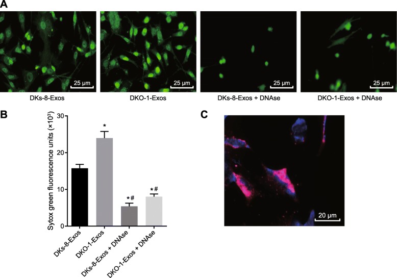
Fig. 4.
Exosomes derived from DKO-1 cells induce NETs formation; a, NETs formation after DKs-8-Exos and DKO-1-Exos were co-cultured with neutrophils (400×); b, fluorescence quantitative assessment of extracellular DNA fluorescence staining. Statistical comparisons were performed using the Mann Whitney U test; c, the interaction of DKO-1 derived exosomes with NETs observed by confocal microscopy (500×). The exosomes are labeled DiD (red) and the NETs are labeled Hoechst 33342 (blue). Neutrophils were isolated and induced to form NETs by stimulation with PMA for 4 h and then incubated with DKO-1-Exos exosomes. * p < 0.05 compared to DKs-8-EXos, and # p < 0.05 compared to DKO-1 Exos. The measurement data was expressed as mean ± standard deviation. Data comparisons between groups were performed using one-way analysis of variance, followed by Tukey’s post hoc test. The experiment repeated three times

